

Introduction
Omphalocele is a congenital anterior abdominal wall defect in which abdominal contents protrude at the base of the umbilical cord into a thin-walled sac.1 The estimated prevalence is approximately 1-3 per 10,000 live births.1–3 Due to the herniation of viscera during fetal development, effective abdominal and chest domains commonly cannot be established.4 Omphaloceles are further characterized by sac size and contents. A giant omphalocele is commonly defined as a defect ≥ 5 cm or one containing at least 50 percent of the liver.5
The management of an omphalocele is individualized and influenced by pulmonary hypoplasia and the extent of visceroabdominal disproportion.6 Traditional approaches for giant omphaloceles involve staged or delayed closure due to the absence of an established abdominal domain. Techniques include silo reduction, elastic dressings, sequential clamping, epithelialization with topical agents, and split-thickness skin grafting over absorbable mesh.5,7
Tissue expansion has been a proposed adjunctive therapy to increase the abdominal domain and enable primary closure for these patients.4,6–8 Tissue expanders (TE) have been described in the subcutaneous or intramuscular spaces of the abdominal wall to enlarge skin or muscle flaps prior to closure, with less frequent use of intra-abdominal expanders to gradually increase abdominal domain.7,9 This case report describes the simultaneous use of intra-abdominal and subcutaneous tissue expansion to achieve primary closure.
This study was granted exemption approval by the University of Mississippi Medical Center Institutional Review Board. Written informed consent for publication of all clinical information and images was obtained from the patient’s parents.
Case Presentation
A 4-year-old female with hydronephrosis and a giant omphalocele was referred to plastic surgery for evaluation and possible closure. She was born with a prenatally diagnosed omphalocele that ruptured at delivery. During her initial hospitalization after birth, a silo was used to reduce the defect, and lateral skin closure was performed. However, the skin closure dehisced, and the wound was subsequently allowed to heal by secondary intention without fascial continuity.
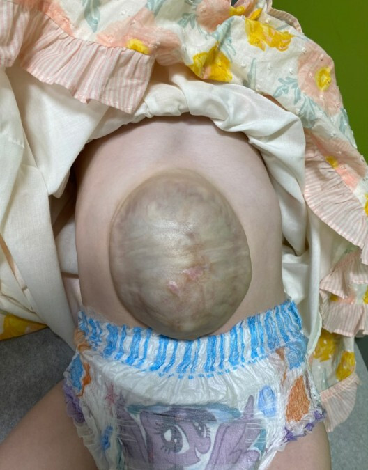
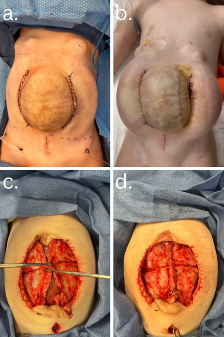
After three years of follow-up, the patient presented to her general pediatric surgeon for elective repair of a large ventral defect containing primarily the left hepatic lobe (Fig. 1). There was presence of epithelialization of the defect without any discomfort or issues with activity or diet. Due to the paucity of healthy skin adjacent to the abdominal wall defect, she was referred to plastic surgery for wound closure with the goal of re-establishing an appropriate abdominal domain. Tissue expansion was offered as a method to increase the soft tissue envelope before hernia repair, enabling the reduction of herniated abdominal contents and primary closure of the fascia and healthy skin edges. Prior to expansion, the abdominal wall defect measured about 7.8 cm transverse diameter and about 12 cm craniocaudal, per the radiologist read on CT abdomen. Two TE (Integra TE Crescent, 5x13 cm, 250 mL capacity) were placed bilaterally in the subcutaneous space along the left and right sides of the omphalocele. A third TE (Integra TE Rectangle, 7x10 cm, 280 mL capacity) was placed in a subfascial, extraperitoneal space inferior to the defect. Blunt dissection was carried onto the thoracic cage where fill ports for each expander were placed (Fig. 2a). The procedure was well-tolerated, and tissue expansion began three weeks postoperatively.
Over the next several weeks, gradual saline inflation was performed. Initial expansion was achieved in the clinic with abdominal TE reaching 135 mL and intra-abdominal expander to 85 mL. After the third visit, the patient was transitioned to anesthesia-assisted expansion due to increased pain and psychosomatic reaction to the filling process. During operative expansion, an additional 35 mL was added to each expander to a total volume of 170 mL in abdominal expanders and 120 mL in the intra-abdominal expander. During this case, the left abdominal expander was noted to have a small area of exposure (Fig. 2b). Given that sufficient tissue had been expanded for reconstruction, the decision was made to proceed with removal and closure the following day.
_tissue_expander_placement_at_the_index.jpg)
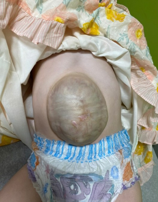
Operative removal of each TE and port was completed without complications. Pediatric surgery then performed extensive lysis of adhesions and reduction of the omphalocele contents into the abdominal cavity. A standard anterior abdominal wall component separation was performed bilaterally. With the components released, the rectus fascia was primarily closed at the midline over the abdominal contents with 0 vicryl figure of 8 sutures. The overlying abdominal skin flaps were further elevated by releasing the lateral portion of the expander capsules, with additional superior and inferior incisions to facilitate tension-free closure of skin (Fig. 2c, 2d). Following closure, the patient’s bladder pressures, and peak airway pressures remained within normal ranges. The postoperative course was uncomplicated, and the patient’s parents report that she is doing well at home with a normal activity level with no restrictions at 1.5 years post-op. No areas of bulging concerning for hernia noted by family or in most recent physical exam documentation (Fig. 3).

Discussion
Giant omphaloceles pose several challenges that limit the success of traditional repair techniques.5,10 Staged silo reduction is commonly used but is limited by the underdevelopment of the abdominal cavity. Similarly, the “paint-and-wait” approach involves using a topical agent to encourage epithelialization but results in a ventral hernia that requires later reconstruction.5,10 These methods address the reduction of viscera but not the underdeveloped abdominal cavity.5,11 Although component separation and mesh reinforcement can aid closure, mesh introduces infection risks, and combined anterior and posterior component separation may weaken the abdominal wall.12–14
This case was influenced by rupture of the omphalocele at birth, which required early silo reduction and healing by secondary intention, resulting in loss of fascial continuity with subsequent epithelialization. This loss contributed to both limited abdominal domain and insufficient adjacent soft tissue at the time of delayed repair. Tissue expansion offers a way to address both soft-tissue deficiency and inadequate abdominal domain.6–9 While tissue expansion itself is not novel,4 the simultaneous use of both intra-abdominal and subcutaneous expanders to address these two constraints concurrently is rarely described in current literature.
Tissue expansion has also not been widely adopted as a primary repair technique for giant omphalocele management due to several limitations. These limitations include the need for multiple procedures, a prolonged treatment course, and higher risk of complications like infection and extrusion.15 Additionally, heterogeneity in technique makes it difficult to establish standardized protocols. Most reports describe tissue expansion as a salvage strategy after other methods have failed, which may reflect hesitancy to pursue a more complex and resource-intensive approach when simpler alternatives may be available.7,10
In our patient, the use of both subfascial and subcutaneous expanders was intentional, as a single plane alone may not have adequately addressed both constraints, particularly given this patient’s significant visceroabdominal disproportion and limited surrounding skin. A pre-peritoneal rectangular TE was used to gradually increase intra-abdominal domain to allow safe visceral reduction, while two subcutaneous crescent-shaped TE on the bilateral flanks recruited additional soft tissue for a tension-free skin closure.4,16 Tissue expansion also promotes the formation of a fibrous capsule around the expanders, facilitating a more controlled and stable closure, with robust blood supply to the skin.17 In this case, the soft tissue and fibrous capsule, along with component separation, allowed for successful closure without the need for mesh.
The rate of expansion is further determined clinically based on patient tolerance and physiologic response. Typically, staged, gradual filling is used to avoid rapid increases in intra-abdominal pressure, either weekly or every other week. In our case, expansion was initially performed in clinic and later under anesthesia due to patient discomfort, with careful monitoring throughout. The main concern with intra-abdominal expansion is elevated intra-abdominal pressure and the risk of abdominal compartment syndrome,9 which can be minimized by gradual expansion. Patients are closely monitored for signs of abdominal compartment syndrome, including respiratory difficulties, elevated peak airway pressures, increased bladder pressure, decreased urine output, and abdominal pain/distention. The authors recommend close monitoring of symptoms for abdominal compartment syndrome throughout the expansion process.
Conclusions
While silastic techniques and topical therapy remain common approaches to giant omphaloceles, tissue expansion provides a reliable alternative by enlarging both the abdominal cavity and soft-tissue envelope. When combined with component separation, primary closure is often achievable. This case report supports the use of simultaneous intra-abdominal and extra-abdominal TE as a promising strategy for managing large omphalocele defects.