History: A four-year-old female with a past medical history of tuberous sclerosis complex (TSC) presented to an outpatient pediatric clinic with a complaint of cough and fever. The patient’s father reported that the patient had had cold-like symptoms for two days. He reported a persistent, non-productive cough with associated rhinorrhea, fever up to 39.7 oC, and one episode of diarrhea. The patient denied otalgia, sore throat, shortness of breath, abdominal pain, vomiting, decreased appetite, recent trauma, and known sick contacts. The father states her TSC was diagnosed three years ago after the patient had a new onset, focal seizure. The patient was begun on levetiracetam, and her seizures have been well-controlled.
Physical Exam: Vital signs were: temperature 37.8oC, pulse 100, respiratory rate 35, and Sp02 93 percent on room air. She weighed 15.4 kg. On examination, the patient was playful and leaning forward slightly while seated next to her father on the examination table. She appeared well-hydrated and well-nourished. Her head, eyes, ear, nose, throat, and abdominal examination were normal. She had tachypnea with the use of abdominal muscles while breathing, and slightly diminished breath sounds bilaterally, without wheezing, rales, or rhonchi. Skin exam revealed several 1-to-2-cm-round hypopigmented macular lesions on her upper and lower extremities.
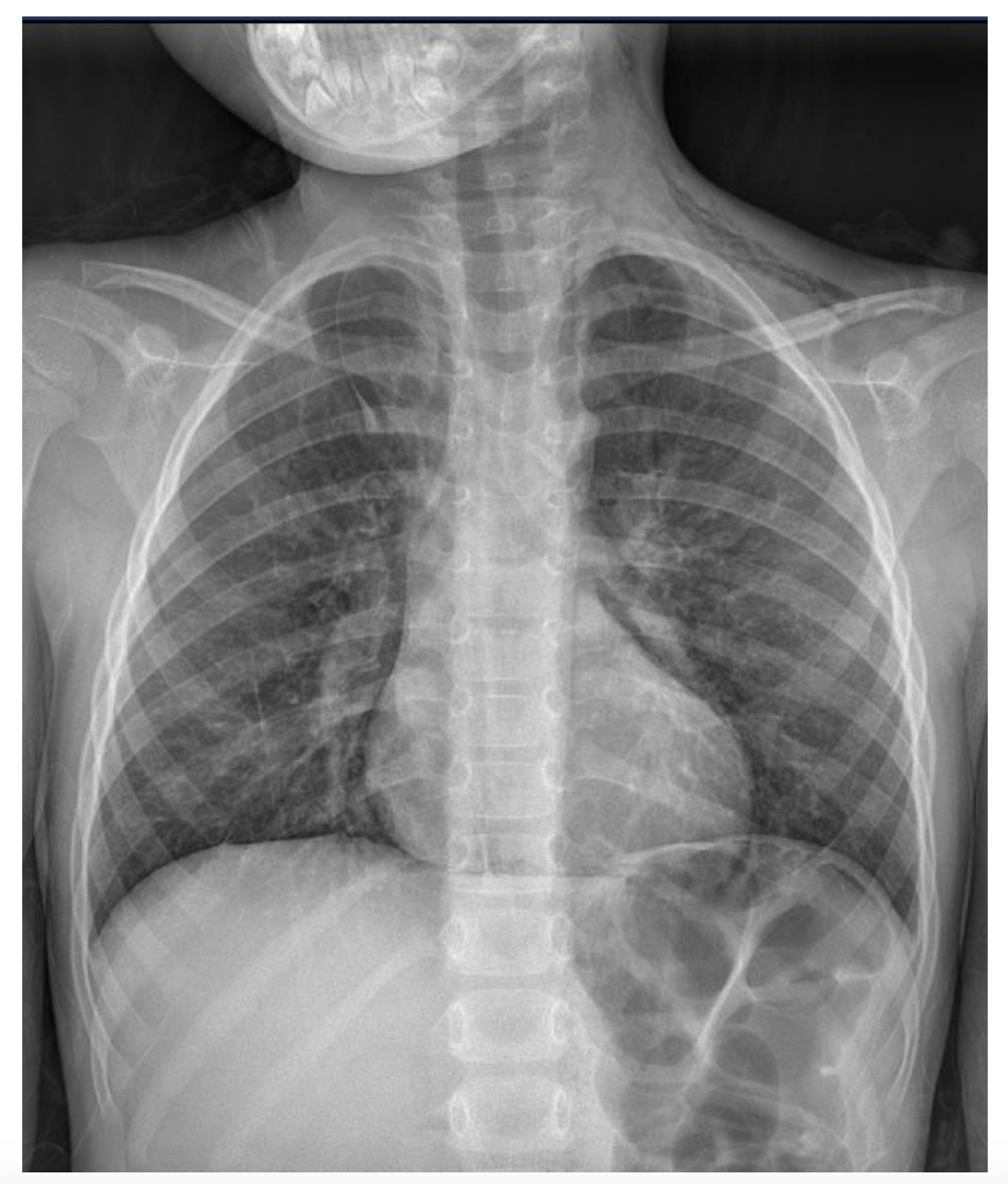
Labs/Diagnostics: A respiratory syncytial virus (RSV) test performed in the clinic was negative. A two-view chest x-ray demonstrated a pneumomediastinum with subcutaneous emphysema within the left neck and supraclavicular fossa, as shown in figure 1. After the discovery of the pneumomediastinum, the patient was directed to the emergency department at a nearby pediatric tertiary care center. A subsequent molecular RSV was positive. The only lab abnormalities noted were a mildly elevated c-reactive protein of 1.6 mg/dL and a slightly low albumin level of 3.6 gr/dL. White blood cell count, hemoglobin, liver transaminases, and all electrolyte levels were normal.

Clinical Course: While in the office, a nebulized treatment of albuterol and ipratropium bromide was administered, and the patient improved clinically, with her oxygen saturation increasing to 95 percent on room air and her accessory muscle use and respiratory rate improving. She appeared in no distress when she left the office. Once admitted to the ED, the patient was placed on 15 L oxygen via a non-rebreather mask and admitted to the pediatric intensive care unit. After a trial on room air, she was discharged to the pediatric ward, where she remained stable. Serial chest x-rays showed improvement in the pneumomediastinum. After two days, the patient was discharged home after a chest x-ray revealed that the pneumomediastinum had almost entirely resolved. At follow-up approximately one week after hospitalization, the patient’s symptoms had completely resolved.
Discussion: SPM occurs when high intra-alveolar pressure results in leakage of air into the pulmonary interstitial space.1 This can be caused by coughing, vomiting, straining, or due to underlying chronic lung disease.2 The incidence varies from 1 in 800 adult patients annually to purportedly 1 in 42,000 pediatric patients. Although SPM is usually benign, complications such as peritonitis, mediastinitis, and tension pneumothorax can arise and warrant urgent evaluation.1
Case studies of RSV resulting in SPM are reported but appear to be rare. However, viral illnesses may not be a direct cause, but rather a cause of coughing that leads to SPM.3
TSC is a genetic neurocutaneous disorder that results in cellular proliferation and migration of benign hamartomas, potentially affecting the brain, heart, skin, eyes, kidneys, and lungs. It is inherited in an autosomal-dominant fashion or as the result of spontaneous mutation. Diagnosis can be made through genetic testing to identify mutations in the TSC1 or TSC2 genes, or it can be made clinically by evidence of two major features or one major feature with two or more minor features.4 TSC affects 1 in 6,000-10,000 people, and is probably underdiagnosed due to the variability of clinical presentations.4,5 Monitoring for disease characterization and progression is needed, including electroencephalogram (EEG), electrocardiogram (EKG), magnetic resonance imaging (MRI) of the brain and abdomen, computed tomography (CT) of the chest, cardiac echocardiography, and subspecialist dermatologic, dental, and ophthalmologic examinations.4
Neurologic complications are common with TSC. The herald of TSC is usually seizure, occurring in as many as 60 percent of cases within the first year of life. The lifetime prevalence of epilepsy is up to 90 percent, but can often present with infantile spasms.5 Brain lesions, including cortical tubers (glioneuronal hamartomas), subependymal nodules, and subependymal giant cell astrocytoma (SEGAs) are common, occurring in up to almost 90 percent of patients.6 TSC- associated neuropsychiatric disorders (TAND) occur in 19.6 to 65 percent of patients and may present as behavioral, intellectual, psychiatric, or psychosocial difficulties.4,6 Although TSC can result in profound intellectual disability in up to 31 percent of cases, approximately 66 percent of patients have a normal intelligence quotient (IQ).6
Skin manifestations occur in up to 97 percent of patients with TSC and are often present at birth.6 Hypomelanotic macules, ash-leaf spots, and confetti macules are also common. Facial angiofibromas, which occur in 25 percent of patients, may be the most specific skin finding for TSC.6 Shagreen patches can also occur.5
Renal manifestations include angiomyolipoma and TSC-related cystic kidney disease which each occur in approximately 50 percent of patients. Renal anomalies can progress to chronic kidney disease or retroperitoneal hemorrhage, necessitating abdominal MRI, glomerular filtration rate estimation, and monitoring of blood pressure.4,6
Cardiac rhabdomyoma can occur in almost 60 percent of patients.6 Rhabdomyomas may be a cause of arrhythmias, flow abnormalities, or heart failure, necessitating periodic monitoring with EKG and echocardiogram. Rhabdomyomas may spontaneously regress; however patients remain at risk for heart failure.4,6
The main pulmonary manifestation of TSC is smooth muscle infiltration of the lungs, or lymphangioleiomyomatosis (LAM), which results in progressive loss of lung function and other lung complications.6 LAM primarily affects females. TSC causes cystic lung changes that can lead to the development of spontaneous pneumothorax.7,8 In fact, as many as 81 percent of females with TSC develop cystic lung changes consistent with LAM.6 Pulmonary complications of LAM-related TSC usually present at the earliest in adolescence.4,8 There is no known association between SPM and TSC.9
Acknowledgments
The authors of this manuscript have no financial interests, activities, relationships, or affiliations that constitute a conflict of interest.
Informed consent for this article was obtained by the patient’s guardian.