Introduction
Leiomyomas are the most common benign uterine tumor affecting 20-80% of women.1–3 The International Federation of Gynecology and Obstetrics (FIGO) classification system stratifies leiomyomas into eight types based on location and percentage of intramural involvement.4 Type 8 leiomyomas, also called parasitic fibroids, are an extremely rare extrauterine variant. Case studies have reported parasitic fibroids attached to the cervix,5 external iliac vessels,6 liver,7 sacral promontory,8 cecum,9 small bowel,10 greater omentum,11 and laparoscopic port sites.12,13 Hypotheses for the etiopathogenesis of parasitic fibroids include arising from type 7 pedunculated fibroids,14 peritoneal metaplasia or de novo myoblast proliferation,8,15 and iatrogenic extrauterine seeding of small leiomyoma fragments from intraperitoneal morcellation during myomectomy or hysterectomy.12,14 To our knowledge no case study has reported findings of a extrauterine leiomyoma seeded during cesarean delivery. Our case highlights a parasitic fibroid found within the preperitoneal tissue of a prior Pfannenstiel incision from a classical cesarean section.
Case Discussion
History, Physical Exam, & Imaging
A 41-year-old G1P1001 female presented with complaints of chronic pelvic pain and intermittent deep dyspareunia. She endorsed iatrogenic secondary amenorrhea with a Mirena intrauterine device (IUD) in situ. Her surgical history includes a prior classical cesarean section. Transvaginal ultrasound showed a 12.8 x 8.7 x 10.4 cm uterus (volume 609.8 cm) with multiple type 5-7 fibroids, the largest of which measured 7.0 x 5.9 x 6.8 cm (volume 147.0 cm). Bilateral ovaries were not visualized due to acoustic shadowing. Central morbid obesity (BMI 38) and a well-healed Pfannenstiel incision were noted on abdominal exam. Bimanual exam revealed a 12-14 week sized multi-fibroid uterus with reproducible pelvic pain upon uterine palpation. She denied a history of abnormal uterine bleeding or cervical dysplasia. Up-to-date pap smear was benign. Patient ultimately desired definitive surgical management via robotic-assisted total laparoscopy hysterectomy (RALTH) with opportunistic bilateral salpingectomy.
RATLH & Mini Laparotomy
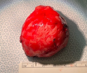
An enlarged asymmetrical multi-fibroid uterus was noted upon laparoscopic entry. Obliteration of the anterior cul-de-sac (OAC) was appreciated with bladder serosa and peritoneum densely adhered to the lower uterine segment and uterine corpus. An isolated 4 cm preperitoneal mass was noted in the right mediolateral anterior cul-de-sac proximal to the bladder (Figure 1). The mass had no appreciable intraperitoneal component. RATLH was then completed via the standard systematic approach for cases involving an OAC.16–18 The surgical specimen comprised of the uterus, cervix, and bilateral fallopian tubes was placed in a 15 cm sterile surgical bag. Due to surgical specimen size and the ability to palpate the preperitoneal mass directly beneath the prior Pfannenstiel incision, the decision was made to proceed with a mini laparotomy. A 6 cm midline incision was made along the previous Pfannenstiel scar, and the deep layers were transected sequentially to the peritoneum, which was entered sharply under direct visualization. The edges of the sterile surgical bag were brought through the mini laparotomy incision. A mini Alexis retractor was placed in the surgical bag and the specimen was removed via minimal myometrial coring (Figure 2). Next, the Alexis retractor was removed, and the tissue capsule of the preperitoneal mass was grasped with Alice clamps and elevated into the operative field (Figure 3). A superficial incision was made over the mass, and it was dissected away from the underlying tissue with Metzenbaum scissors. The mass appeared consistent with a parasitic leiomyoma and was sent to pathology in a separate bag (Figure 4). Next, the mini laparotomy was closed in standard fashion. Lastly, the da Vinci robot was redocked and the vaginal cuff and peritoneal portion of the mini laparotomy incision were closed with running V-Loc suture. No complications were noted. Estimated blood loss was < 100 mL.
Pathology Findings
Pathology reported a 15.8 x 14.9 x 8.3 cm markedly asymmetrical uterus weighing 571g. Numerous subserosal and intramural leiomyomas were noted, the largest measuring 7 cm. Histology revealed benign fallopian tubes, inactive endometrium with exogenous progesterone effect, benign cervix with chronic cervicitis, and an IUD. The preperitoneal lesion was reported as a 4.5 x 3.5 x 2 cm partially encapsulated benign leiomyoma weighing 41g.
Discussion
Different hypotheses exist regarding the etiopathogenesis of parasitic fibroids. Extrauterine leiomyomas have been reported in patients without a history of pelvic surgery.6,11,19 However, most parasitic fibroids are due to iatrogenic seeding of small leiomyoma fragments during gynecologic surgery.5,7–10,12–14 Parasitic fibroids have occurred following both laparoscopic and abdominal myomectomy and hysterectomy.5,7–10,12–14 Many cases have involved morcellation,8,12,14,20 which can generate small fragments of leiomyoma tissue that can seed into extrauterine tissue and arise as a parasitic fibroid. The incidence of parasitic fibroids after laparoscopic morcellation is between 0.95 to 1.2%,14,20 supporting the hypothesis that most parasitic fibroids are iatrogenic.
Although type 8 leiomyomas may arise from peritoneal metaplasia or de novo myoblast proliferation,8,15 the parasitic fibroid in our case likely arose from iatrogenic seeding of leiomyoma fragments during a classical cesarean section. Classical uterine incisions are midline incisions through the contractile portion of the myometrium. Incising the myometrium has the theoretical potential to create myoma debris that can then seed into extrauterine tissue. This case is unique for two reasons. First, it is the only case to our knowledge to report seeding of a parasitic fibroid during cesarean delivery. Second, almost all type 8 leiomyomas occur within the intraabdominal cavity, with 93% occurring within the pelvis.21 However, this case involves a preperitoneal lesion within a prior Pfannenstiel incision.
Parasitic fibroids remain an extremely rare variant of leiomyomas with approximately 30 reported cases in the literature.22Although the majority of cases have been caused by intraabdominal morcellation,8,12,14,20 morcellation of gynecologic tissue is no longer routinely performed due to the risk of intraperitoneal dissemination of cancerous cells.23 Using endoscopic bags when removing surgical specimens and employing copious irrigation during cases that disrupt fibroid or myometrial tissue can likely decrease the risk of parasitic fibroids. It is important for surgeons to be knowledgeable on the etiopathogenesis of parasitic fibroids and to be mindful of potential seeding during gynecologic and obstetric procedures.
Informed Consent
Written consent has been obtained from the patient for publication of this case report.


