Introduction
Low back pain is a common complaint that can stem from various etiologies, ranging from musculoskeletal issues to serious neurological conditions. Primary care physicians hold a crucial role in evaluating patients with persistent symptoms and considering rare diagnoses when faced with atypical or prolonged presentations of common complaints. In the following case report, we discuss a case of clear cell meningioma, a rare spinal tumor, as the uncommon etiology of this common presenting symptom. We prepared this manuscript using the CARE Guidelines.1
Case
A woman in her 20s presented for a new patient visit at a primary care clinic and reported a two-year history of lower back pain. Her past medical history included obesity, generalized anxiety disorder, and seasonal allergies. She reported pain initially started in the lumbrosacral region then progressed with radiating to her bilateral hips. She denied preceding trauma, fever, unintentional weight loss, fever, intravenous substance use, changes in bowel or bladder function, lower extremity weakness, or sensory deficits. Of note, imaging obtained in the emergency department (ED) two years prior included x-ray of the bilateral hips and computed tomography of the lumbar spine, both interpreted as normal. Soon after this ED visit, she was evaluated by sports medicine who recommended MRI, but she could not obtain imaging due to uninsured status. At the time of presentation to primary care, she had started new employment and gained health insurance coverage. She reported pain worsened the past 6 months and limited her ability to perform job-related tasks. Non-steroidal anti-inflammatory medications and home exercise had not provided symptom relief.
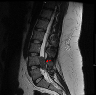
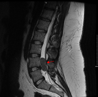
On examination, the patient demonstrated mild diffuse tenderness in the lumbar region. Neurologic exam including gait and reflexes was within normal limits. Given the chronicity of her symptoms, further imaging was indicated to evaluate for an underlying etiology of her pain. The patient underwent an MRI of the lumbar spine, which revealed a well-circumscribed mass noted in the spinal canal at the level of L4-L5. This mass occupied the entire spinal canal and compressed the adjacent neural structures. The lesion appeared intradural, and initial differential diagnosis included meningioma, schwannoma, ependymoma or other primary central nervous system (CNS) neoplasm. Additional cross-sectional imaging of the chest, abdomen, and pelvis did not show evidence of metastasis.
Following the diagnosis, the patient was referred to a Neurosurgery team at a tertiary medical center for further management. Her neurosurgery team proceed with surgical resection of the tumor, which was performed successfully with no immediate complications. Pathological examination confirmed the diagnosis of clear cell meningioma. Adjuvant radiation therapy was considered to minimize the risk of recurrence but deferred due to potential side effects. Fortunately, continued surveillance with imaging and clinical examination showed no signs of recurrence as of 18 months following surgical resection.
Discussion
Guidelines from the American Pain Society highlight the importance of a thorough history and physical examination, providing reassurance, initial pain management, and physical therapy while avoiding routine imaging for patients with nonspecific low back pain (LBP).2 If symptoms persist despite 6 weeks of conservative therapy, the American College of Radiology (ACR) appropriateness criteria recommends MRI lumbar spine without IV contrast as the most appropriate imaging modality.3 Intradural tumors and spinal cord abnormalities are often inadequately visualized on CT scans, as noted in this patient case in which CT did not detect signs of a tumor. Referencing ACR guidelines in clinical documentation may prove beneficial in cases where radiology prior authorization is requested.
Primary spinal cord tumors account for only 2-4% of all primary CNS tumors.4 Spinal meningiomas typically occur in patients over age 70, with a four-fold greater incidence in women as compared to men.5 Clear cell meningiomas are a rare CNS tumor that may present with atypical symptoms when located in less common areas. The majority of clear cell meningiomas are intracranial, with only a small margin being found in the spine.6 Clinical presentation varies based on location of tumor and on the depth of the tumor within the cord. Clear cell meningiomas in the spine may produce neurologic deficits and pain due to local compression of the spinal cord, vessels, nerves, and adjacent structures.4 Overall, the most common presenting symptoms of spinal meningiomas are motor dysfunction (92%), sensory disturbance (78%), pain (76%), gait disturbance (42%), and bowel or bladder dysfunction (28%).6 MRI often shows a well demarcated, intradural, homogeneously enhancing mass. Surgical resection is the gold standard treatment for spinal meningiomas, often alleviating symptoms with minimal risk of recurrence postoperatively.7 Radiation therapy is a consideration for higher grade tumors or in patients unable to undergo surgery, though the role of non-surgical therapies is variable.
This case of a young woman with low back pain ultimately diagnosed with clear cell meningioma underscores the profound impact of uninsured status on the timely diagnosis and management of serious health conditions. Prior research demonstrates that racial minorities and patients with lower socioeconomic status have worse survival outcomes when diagnosed with CNS tumors.8 Primary care physicians hold a crucial role in advocating for equitable access for all patients and reducing healthcare disparities on a system level.
Acknowledgements
The patient graciously provided her consent for sharing details and lessons of her case in the medical literature. The authors have no conflicts of interest to report.