


Thromboembolism is the most common cause of acute upper extremity ischemia.1 There are multiple underlying conditions that can result in thromboembolism. The following are some common disease processes that lead to limb ischemia: atrial fibrillation, valvular heart disease, isolated ischemic heart disease, left atrial myxoma, left ventricular aneurysm, penetrating injuries, arterial access complications, or valvular vegetations in infective endocarditis.2 Uncommonly, inadvertent intraarterial drug injection is also a cause of thromboembolism related to acute digital ischemia.3 In the following case report, we discuss a case of inadvertent intraarterial drug injection in a patient with a long history of intravenous drug use. We prepared this manuscript using the CARE Guidelines (for CAse REports).4
CASE
A 33-year-old female with a history of intravenous drug injection presented to an urgent care clinic with a chief complaint of discoloration and decreased sensation to the right index finger. The patient reported a 10-year history of intravenous drug use. Eight days prior, she injected fentanyl into her wrist, near the anatomical location of the radial artery. Soon after injecting, she began having a bluish discoloration in her right thumb, which lasted approximately one day and spontaneously resolved. Approximately four days after the previous injection, she injected the same location again. She reports later that day, her right index finger turned blue, with associated decreased sensation and coolness to touch. After a total of four days without improvement, she presented to an urgent care clinic.
She denied fever, weight loss, night sweats, chronic cough, or previous issues with abnormal bruising. She denied past infections from intravenous drug use, other than Hepatitis C, and denied use of contaminated needles. She admitted to frequent intravenous drug use with difficulty finding veins due to her long history of drug injection.
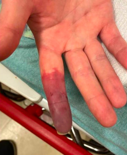
On initial exam, she was anxious appearing with normal vital signs. Her right index finger was noted to be cyanotic and cool to the touch. She had lack of sensation distally in the index finger but did have full active range of motion. The cyanosis began at the mid proximal phalanx and increased distally along the finger. She was noted to have numerous skin changes over both arms and hands associated with frequent injections. She had a small, crusted lesion to the dorsum of her right thumb which appeared to be a lesion from previous injection. In addition, her left hand was mildly edematous up to her mid forearm. See Figure 1 and Figure 2 for Urgent Care initial imaging revealing mottling to right hand and discoloration to right index finger.
The urgent care nurse practitioner discussed the patient’s case with an orthopedic hand surgeon and emergency medicine colleague, who recommended the patient be seen at the local Emergency Department immediately for evaluation and treatment. The patient presented to the Emergency Department the same day. The patient’s chief complaint was as above, with the addition of skin color changes to both hands and forearms.
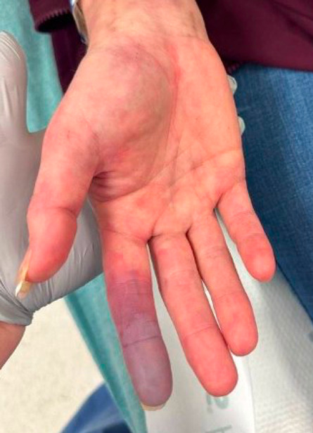
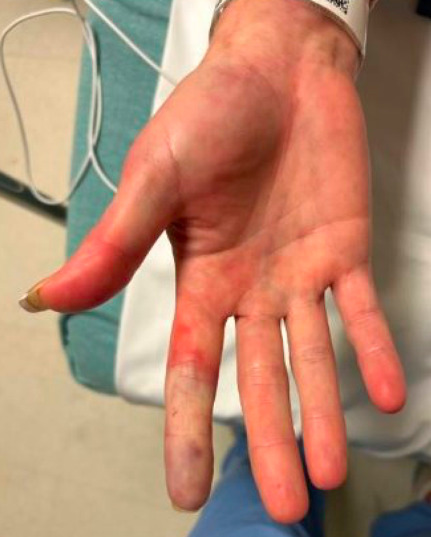
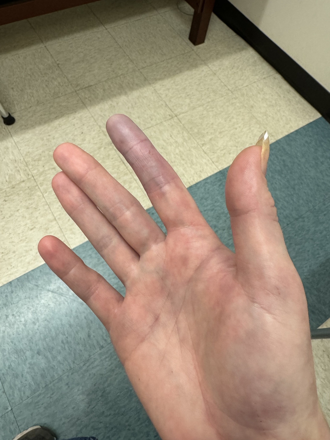
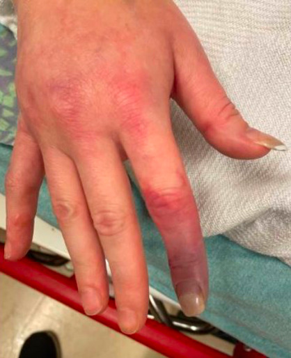
On physical exam, the patient was initially hypertensive to 182/114, afebrile at 98.2, tachycardic at 119 bpm, with a normal respiratory rate and normal oxygen saturation. The patient presented with significant discoloration of the right index finger, circumferentially, from the distal phalange to the metacarpophalangeal joint; there was also palpable coolness to the finger, as well as decreased capillary refill. There was scattered mottling of the tissue of both hands and both forearms, circumferentially. She had decreased light touch sensation to her right index finger. The right radial pulse was palpable. The remainder of her physical exam was benign. See Figure 3 for a palmar image of the right hand showing discoloration as described above; likewise, see Figure 4 for a dorsal image of the right hand showing discoloration as described above.


The initial consults placed from the Emergency Department included: Orthopedic Surgery, Vascular Surgery, and Internal Medicine.
The results of the initial ED workup are as follows:
White blood count 8.6
Hemoglobin 12.7
Hematocrit 38.0
Mean Corpuscular Volume 92.5
Platelet count 301
Creatinine 0.66
Blood Urea Nitrogen 9.6
Sodium 137
Potassium 3.8
Chloride 104
CO2 21 (low)
Partial Thromboplastin Time 21.1 (low)
International Normalized Ratio 1.06
Erythrocyte Sedimentation Rate 9.0
C-reactive Protein <0.3
CTA imaging was without concern for acute vessel occlusion in bilateral upper extremities. Specifically, the CTA visualized widely patent brachial arteries, radial arteries, and ulnar arteries with preserved flow to the hands.
The patient was evaluated by a vascular surgeon who recommended the initiation of a heparin infusion, obtaining blood cultures, and covering empirically with vancomycin and unasyn for prophylactic endocarditis coverage.
The patient was admitted to Internal Medicine for vascular compromise of the right index finger, as well as concern for sepsis secondary to cellulitis with suspected bacteremia vs endocarditis. The inpatient team modified the antibiotic regimen from unasyn to cefepime; vancomycin was continued. The heparin infusion was continued, as well.
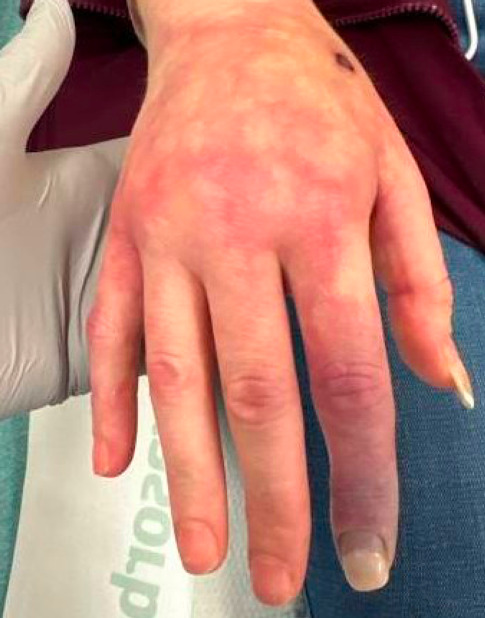
On hospital day two, the patient underwent a formal transthoracic echocardiogram which revealed the following: normal left ventricle size and function, ejection fraction of 60%, and no valvular disease. Heparin, vancomycin, and cefepime were continued, and the physical exam improved. She remained hemodynamically normal and the blood cultures remained negative. Surgical specialties continued to recommend conservative management. See Figure 5 and Figure 6 for day two imaging; figure 5 shows evolving color change on the dorsum of the right hand, and figure 6 shows evolving color change on the palmar surface of the right hand.


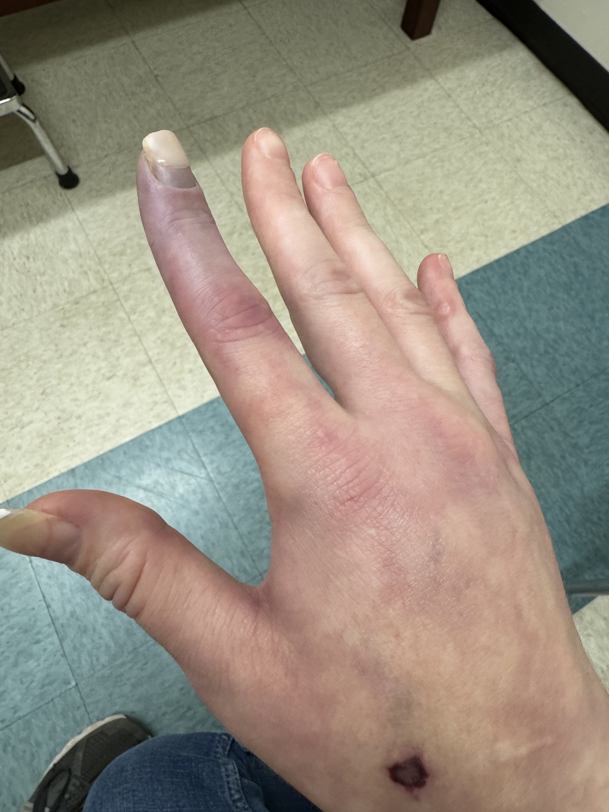
On hospital day three, the patient’s antibiotic regimen was transitioned from cefepime and vancomycin to doxycycline (100mg orally twice daily x 6 days) and augmentin (875-125mg orally twice daily x 6 days). She was also transitioned from the heparin infusion to Eliquis (5mg orally with a tapered schedule). Her physical exam continued to improve, and her hemodynamics remained stable. She reported complete resolution of her previous light touch sensation deficit. The Internal Medicine team deemed the patient stable for discharge home with the above regimen, and close outpatient follow-up with Vascular Surgery, Orthopedic Surgery, and a Primary Care Physician. See Figure 7 and Figure 8 for day-of-discharge imaging; Figures 7 and 8 are palmar and dorsal images of the right hand, respectively, that show improved mottling of soft tissues of the hand, but evolution of soft tissue change to the right index finger. There was no further documentation of any outpatient follow up.
DISCUSSION
Providers are taught the “6 P’s of Ischemia”: pain, pallor, poikilothermia, pulselessness, paresthesia, paralysis.5 Identifying acute ischemia related to intraarterial injection can be difficult, as other diagnoses may overlap with findings.6 Patients are likely hesitant to disclose the use of intravenous drugs, leading to delayed diagnosis and treatment.7 Prompt diagnosis leads to prompt treatment which ultimately improves morbidity. Most commonly, intra-arterial injection of drugs causes direct vessel trauma, arterial spasm, local toxicity, or embolism of matter. Acute limb ischemia is a clinical diagnosis, although imaging may be warranted for further work-up. Treatment varies based on severity and location of thrombosis.8