

- Androgenetic alopecia (AGA) is the most common form of hair loss globally.1 AGA is chronic and influenced by both genetics and environment. In men, AGA presents as a receding frontal hairline beginning as bitemporal thinning of the frontal scalp propagating towards the vertex, whereas in women, diffuse hair thinning between the frontal scalp and vertex with frontal hairline retention is seen, as in Figure 1. Topical minoxidil and oral finasteride are FDA-approved, but several non-approved treatments such as platelet-rich plasma (PRP), low-level light therapy, micro-needling, and nutrient supplementation have shown efficacy.2

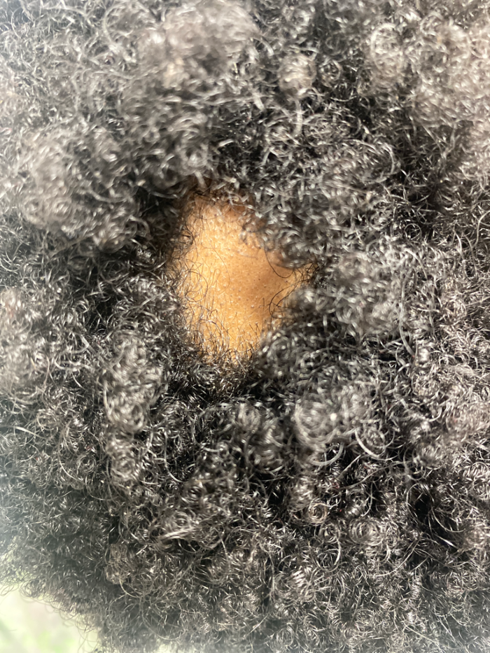
- Alopecia areata (AA) is a chronic autoimmune hair loss disease that affects 2% of the population.3 AA has a diverse presentation, ranging from localized patches (see Figure 2) to diffuse or total hair loss of the scalp (alopecia totalis) to entire body hair loss (alopecia universalis). Pathogenesis is not fully understood but believed to be due to the collapse of immune privilege within hair follicles. Traditionally, the driver of the disease has been described as T-helper (Th) 1/ interferon skewing, treated with topical and intralesional steroid injections and broad immunosuppressants. Recent studies suggest additional mediators including the Th2 pathway, interleukin (IL)-9, IL-23, and IL-32 as contributors and potential therapeutic targets.4 Emerging drugs, including Janus kinase (JAK) inhibitors like barcitinib and ritlectinib, show promise with recent FDA approval.

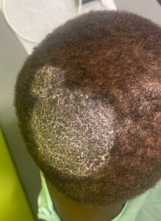
- Central centrifugal cicatricial alopecia (CCCA) is a scarring alopecia predominantly affecting women of African descent, marked by permanent hair loss on the scalp’s vertex or crown with centrifugal spread, as in Figure 3.5 While once attributed solely to hairstyling practice, recent research reveals contributors including genetic variants and systemic conditions. Initial managements target eradicating damaging practices, while steroids and antibiotics combat inflammation and potential infection. Minoxidil may stimulate hair growth in unaffected areas. Emerging treatments include metformin and PRP. Hair transplantation is considered for stable cases, and camouflage techniques like wigs aid advanced stages. Understanding CCCA’s complexity informs comprehensive management approaches.6

- Tinea capitis is a highly contagious fungal scalp infection.7 In the United States, it is caused most commonly by Trichophyton tonsurans. It varies in presentation from scaly dermatosis (see Figure 4) to inflammatory lesions known as kerion.8 Diagnosis involves clinical examination, KOH analysis, and fungal culture. Prompt treatment, combining oral antifungals with topical agents and selenium sulfide shampoo, is crucial to prevent scarring and hair loss. In severe cases, oral corticosteroids may be added to reduce inflammation.

- Telogen effluvium occurs when a significant number of hair follicles are prematurely pushed into the telogen (resting) phase of the hair growth cycle. Triggers such as physical or emotional stress, illness, certain medications, nutritional deficiencies, or hormonal changes can cause this response.9 Diagnosis is based on clinical presentation (see Figure 5) while a hair pull test provides confirmation. In most cases, telogen effluvium is a self-limiting, non-scarring condition, and the hair regrows within 3-6 months.10 Treatment involves managing the underlying cause, ensuring adequate nutrition, and topical minoxidil.

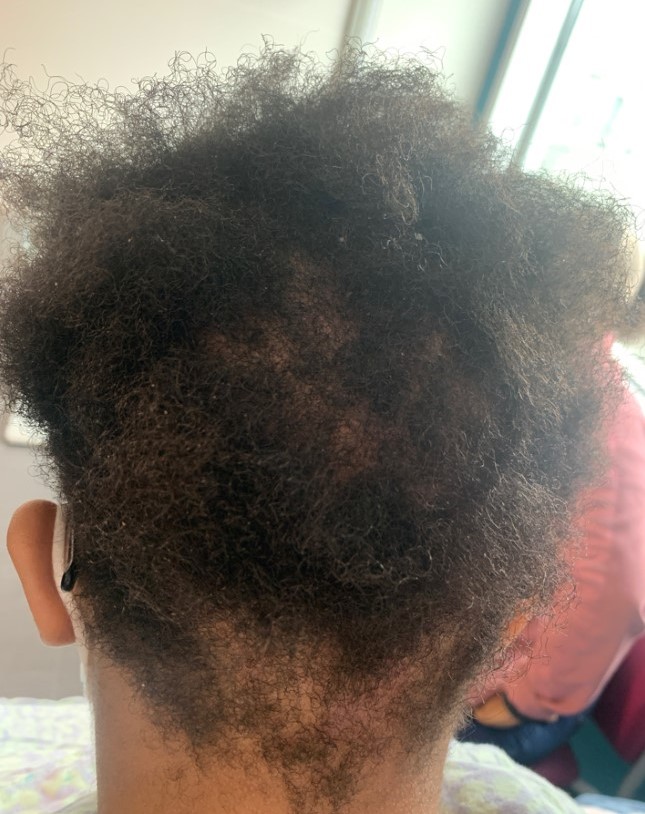
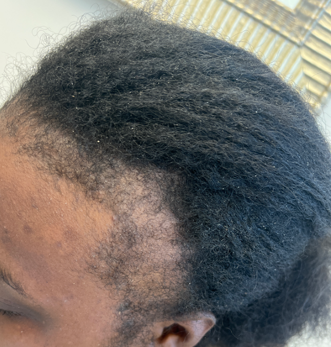
- Traction alopecia is a preventable form of hair loss caused by repetitive or prolonged tension on the hair, often from tight hairstyles like braids, ponytails, and cornrows.11 It is associated with professions like ballet and gymnastics due to the expected hairstyles, and it also most commonly affects women of African descent.12 Early signs include thinning at the frontal hairline (see Figure 6), progressing to “fringe sign” where fine hairs remain along the edges of the original hairline and eventually scarring and irreversible hair loss. Treatment involves discontinuing tight styles promptly, using topical corticosteroids, and potentially incorporating minoxidil or biotin supplements.

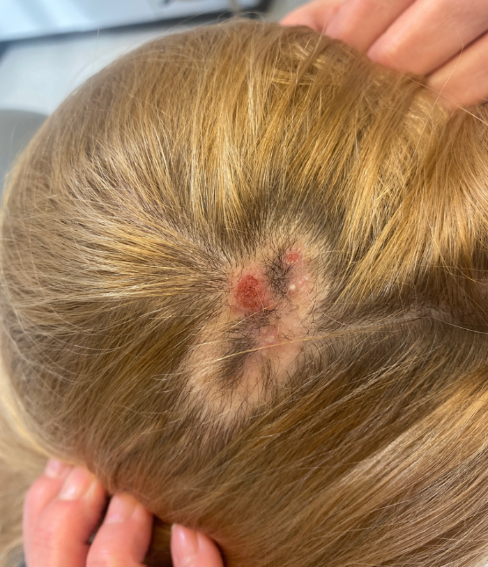
- Trichotillomania is characterized by the urge to pull out one’s own hair, leading to hair loss (see Figure 7) and significant distress or impairment in daily life.13 It is linked with obsessive-compulsive and anxiety disorders and is more prevalent in women.14 Effective treatment involves a combination of cognitive-behavioral therapy and sometimes the use of selective serotonin reuptake inhibitors or antipsychotics. Early recognition and intervention are crucial to prevent the development of chronic, disfiguring hair loss and associated psychological distress.

-
Discoid lupus erythematosus (DLE) primarily affects the scalp, face, and ears and is the most common form of cutaneous lupus erythematosus.15 Initially, lesions appear as purplish macules or papules, evolving into discoid or coin-shaped plaques with peripheral hyperpigmentation. A significant complication of DLE is scarring alopecia, where hair follicles become clogged by adherent scales. Affected areas may develop atrophic lesions with peripheral discoloration and central depigmentation leading to permanent follicular damage. First-line treatments include photoprotection, topical or intralesional corticosteroids, and topical calcineurin inhibitors.
-
The hair pull test is a simple yet valuable diagnostic tool for evaluating hair loss causes.16 Active shedding is indicated by more than 10% of hair coming out when gently pulling on a small bundle of scalp hair. This test helps differentiate conditions like telogen effluvium, androgenetic alopecia, and alopecia areata. However, the test is subjective and has limitations - it does not definitively rule out hair loss.17 Physicians should use the hair pull test alongside other diagnostic methods to arrive at an accurate diagnosis and treatment plan.
-
An escalating prevalence of invasive fungal infections (IFIs) has been seen globally, attributed to a growing population of immunocompromised patients.18 While culture-based methods remain the gold standard for diagnosing IFIs, nonculture diagnostics like serological assays and molecular techniques offer faster and more sensitive results.19 However, none of these methods can replace culture entirely; these methods should be used alongside host assessments and radiographic features for optimal patient management. Despite advancements, challenges persist in IFI diagnosis, necessitating improved stewardship, cost-effectiveness, and accessibility of fungal diagnostics.